


This disease is poorly understood, although several thousand observations with diagnosis and follow-up have been described.
The diversity and non-specification of the clinical picture of small pelvic varicose veins leads to gross errors of diagnosis, which will affect the results in the future.
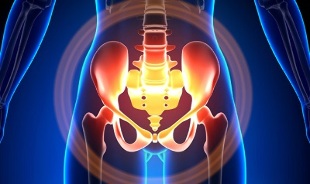
Characteristics of small pelvic varicose veins
The pelvic veins are several times longer than the arteries, leading to their greater capacity. This is due to the phylogeny of the vascular system of the pelvic region. The pelvic veins are highly adaptable and potentially prone to renewal, contributing to the formation of a tightly tangled network.
The speed and direction of blood circulation is regulated by valves controlled by complex humoral mechanisms. The valves balance the pressure in different parts of the venous network.
When the valves stop performing their functions, blood stagnation develops, which leads to vascular pathology and the formation of varicose veins. The uniqueness of pelvic veins lies in the fact that the wide ligaments of the uterus, which are wide of the vessel lumen, can narrow, leading to pathology.
Causes of occurrence
Abnormal enlargement of the pelvic veins can be due to the following reasons:
- Disruption of the bloodstream;
- Deviation of the Vienna trunk;
- Compression of collaterals in a modified position of the uterus, for example, by retroflection;
- ovarian venous valve insufficiency (congenital or acquired);
- obstructive post-phlebitis syndrome;
- connective tissue pathology;
- arteriovenous angiodiplasia;
- long sitting, hard physical labor;
- varicose veins of the lower extremities;
- Pregnancy (3 or more) and childbirth (2 or more);
- Diseases of the female genitalia (chronic salpingo-oophoritis, ovarian tumors, uterine myoma and genital endometriosis);
- Adhesion of pelvic organs;
- Obesity.
Classification according to the degree of the disease
The following degrees are distinguished according to the size of the varicose vein:
- up to 0. 5 cm, "corpus" course of ships;
- 0, 6-1 cm;
- more than 1 cm.
Disease variants
- Varicose veins of the perineum and the vestibule of the vagina;
- Small pelvic venous edema syndrome;
Symptoms
- The most common - frequent pain in the lower abdomen after prolonged static and dynamic stress in the perineum. The pain intensifies in the second phase of the cycle, after hypothermia, fatigue, stress, exacerbation of various diseases.
- Feeling of "inappropriate place", pain during and after sex. Dysmenorrhea - a disorder of the menstrual cycle, including pain.
- Abnormal secretion of gonads.
- Blood stagnation leads to infertility, miscarriage, abortion.
- Disorder of urine due to dilation of bladder veins.
Diagnostics
Diagnosis of the disease with complaints alone is successful in only 10% of cases.
Palpation of the inner walls of the pelvis allows you to feel the elongated rings and venous nodes. Cyanosis of the vaginal mucosa can be seen when looking in a mirror.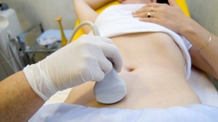
The procedure of choice is an ultrasound examination with a color Doppler map, which allows you to detect not only varicose ovarian veins, but also venous thrombosis, post-thrombophlebitis. Ultrasound shows tortuosity, "worm-like", structures without signal reflected, localized on the lateral surface of the uterus.
The Doppler effect is based on the blue and red tones of the venous and arterial circulation.
Ultrasound examination device with the help of a special program detects the movement of blood from the sensor and in another direction, calculates the speed of blood flow and the type of vessel.
But the exact definition of a vein or artery remains with the doctor. The Doppler method works in almost all cases, the exception to the rules is dictated by our body, since the blood from the heart is not always arterial and vice versa.
Thus, the ultrasound doctor sees this arterial or venous vessel, its size, the frequency of blood flow in it, and many indicators that are not necessary for the average person, but play an important role in the diagnosis. Transabdominal and transvaginal sensors are used for this purpose.
5. In 7% of cases the disease is recognized at the time of screening. Usually the diameter of the ovary vein is 0. 4 cm.
CT and MRI are very accurate. With these methods it is possible to detect the accumulation of varicose veins around the uterus, ovaries and these organs. It is possible to identify concomitant pathology.
Phlebographic research is a very reliable method.
Contrast method is performed at the height of the Valsalva test, against blood flow. This allows you to accurately see the valve failure.
Left X-ray radioscopy, renal phlebography, super-selective phlebovarioscopy, and phlebovariography are also used on both sides. These methods make it possible to determine the hemodynamic and anatomical changes in the renal veins and the areas where the gonadal veins enter them.
Super-selective phlebovarioscopy is performed by catheterization of the gonadal veins through the contralateral femur or subcutaneous vein, followed by contrast injection.
Most blood from the varicose veins of the vascular plexus is taken into the ovarian vein. But under conditions of hypertension, it occurs through extrauterine uterine veins into the iliac vein. The venous plexus through which drainage can occur includes the sacral and bladder plexuses.
Left-sided phlebovarography reveals 3 stages of venous stasis in the left ovary uviform plexus:
- There is no drain from the left ovary, or it follows an extra short path.
- There is an extra long way to go.
- Two additional drain paths are visible, or one additional and auxiliary.
Stages 2 and 3 produce varicose veins of the right ovarian plexus.
Laparoscopy is used for differential diagnosis. Abnormally twisted veins are located in the ovarian region, in the direction of the round and wide yokes. They look like large cyanotic conglomerates, with a thin and tense wall.
The difficulty of diagnosis lies in the fact that the disease is often hidden behind the signs of the inflammatory process, differs in clinical manifestations, is covered in the form of endometriosis, prolapse of internal organs, postoperative neuropathies and many extragenital diseases.
Treatment
The main goal of treatment is to relieve venous reflux. Conservative treatment is used in the early stages of the disease. In the later stages of the disease, surgery is the treatment of choice.
Conservative treatment

It involves normalizing venous tone, improving hemodynamics and trophic processes.
Symptomatic treatment of individual symptoms. Nonsteroidal anti-inflammatory drugs for pain, bleeding - hemostatic therapy.
The mainstay of conservative treatment is venotonic drugs and platelets.
Phlebotonics - improves the tone of the blood vessel wall and increases blood flow. With this disease, it is best to consult a gynecologist about certain medications.
Physiotherapy is an important method.
Surgical treatment
- Resection of varicose veins.
- Gonadal cavitation.
- Sclerosis in laparoscopy.
- Ovarian vein occlusion using X-ray endovascular methods.
Folk remedies
Since the main factor in the onset of the disease is the weakness of the valvular apparatus, for this pathology are also used all the folk remedies used for varicose veins of the lower extremities.
Most commonly used: common hazelnut, hops, nettle, horse chestnut, dandelion root, kombucha, willow, oak, St. John's wort, string, pollen and many other plants.
Effective: oak, chestnut, willow, chamomile, pharmacy, dried herbs, St. John's wort, string baths treatment.
Prevention
- The first thing you should do if you have any of the above complaints, prognosis or diseases is to contact your gynecologist.
- It is necessary to normalize the work regime and rest, try not to stay in a vertical position for a long time, physical overload.
- Do exercises "pedal", "birch stand", "scissors legs", follow a diet for prevention: Eat foods rich in vitamins E, P, C, try to eat only white meat, low-fat meat, replace it with fruits, vegetables.
- Drink plenty of fluids, but not less than 1. 5 liters per day.
- Get rid of excess weight, bad habits.
- Ask your doctor about wearing a compression garment, this will improve blood flow from the lower extremities, thus reducing swelling in the pelvis.
- Avoid baths, saunas, steam rooms, hot baths.
In order not to get sick with such a difficult diagnosis, you should follow the prophylactic recommendations listed above. Treat your health as the most precious thing in life.
For the slightest suspicious symptoms, from which you will not be able to escape in a few days, you should consult a doctor. He must provide you with highly qualified help and save you from suffering.